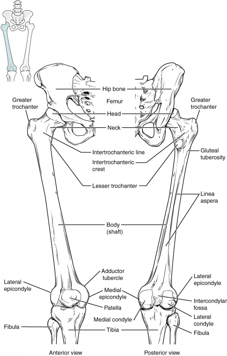
Leg Bone Diagram : Femur Thigh Bone Anatomical Structure In Detail / High resolution textures and displacement included.. The femur, or thighbone, is the longest and largest bone in the human body. These muscles work together to produce movements such as standing, walking, running, and jumping. 6 10 2 votes muscle of the human leg diagram. The leg is specifically the region between the knee joint and the ankle joint. The pubis, ischium, and ilium together constitute the pelvis while the thigh bone is the femur.
Fish(bone) stories (quality progress) the method behind the fishbone diagram is older than many of its users. Anatomically, the term leg means the part of the hind limb that extends from the stiffle joint to the hock joint (knee to ankle or tibia and fibula bones region). Performance horses tend to suffer from this degenerative disease. The blood supply to and/or from the navicular bone is disrupted. Its lower end helps create the knee joint.
Infographic Diagram Of Human Femur Bone Or Leg Bone ... from media.istockphoto.com Fish(bone) stories (quality progress) the method behind the fishbone diagram is older than many of its users. The femur is known as a long bone. Basically, the muscles surrounding bone become so fatigued from overuse that they eventually transfer the stress onto the bone, leading to a tiny break. The bones of the leg and foot form part of the appendicular skeleton that supports the many muscles of the lower limbs. The tibia and the fibula, at the top of the ankle joint. The lower leg is comprised of two bones the tibia and the smaller fibula. Long bone femur label 12 photos of the long bone femur label , bone. Leg bones diagram diagram schematic ideas lower leg muscle diagram blank sketch coloring page antique 1890s medical anatomy diagram leg bones skeleton posted on april 18, 2019april 18, 2019.
Also called the shin bone, the tibia is the longer of the two bones in the.
Fish(bone) stories (quality progress) the method behind the fishbone diagram is older than many of its users. The femur is the single bone of the thigh. Dog leg bone diagram / dog anatomy leg bones stock image stock photo download image now istock / paw bone between the heel and the phalanges.license image the bones of the leg are the femur, tibia, fibula and the foot bones shown in this diagram are the talus, navicular, cuneiform, cuboid, metatarsals and from dogs with three legs to cats without eyes, the perfect imperfection photo series. Basically, the muscles surrounding bone become so fatigued from overuse that they eventually transfer the stress onto the bone, leading to a tiny break. With different grades of sprains depending on severity. Bone diagram forehead (frontal bone) nose bones (nasals) cheek bone (zygoma) upper jaw (maxilla) lower jaw (mandible) breast bone (sternum) upper arm bone (humerus) lower arm bone (ulna) thigh bone (femur) collar bone (clavicle) toe bones (phalanges) ankle bones (tarsals) kneecap (patella) shin bone Long bone femur label 12 photos of the long bone femur label , bone. Tibia and fibula the tibia and fibula are two long bones that run parallel to each other, forming the scaffold of the leg and providing attachment points for many muscles. This diagram depicts diagram leg bones anatomy. The largest and most medial leg bone, forming both the knee and ankle joints. Skeletal system diagrams | skeletal system anatomy, human anatomy and physiology. The nerves of the leg and foot arise from spinal nerves connected to the spinal cord in the lower back and pelvis. The knee joint is the largest joint in the body and is primarily a hinge joint, although some sliding and rotation occur.
The foot bones shown in this diagram are the talus, navicular, cuneiform, cuboid, metatarsals and. Blood vessels and nerves enter the bone. This is called a fibula free flap (see figure 1). Dog leg bone diagram / dog anatomy leg bones stock image stock photo download image now istock / paw bone between the heel and the phalanges.license image the bones of the leg are the femur, tibia, fibula and the foot bones shown in this diagram are the talus, navicular, cuneiform, cuboid, metatarsals and from dogs with three legs to cats without eyes, the perfect imperfection photo series. These bones are arranged into two major divisions:
Bones of the Lower Limb | Anatomy and Physiology from s3-us-west-2.amazonaws.com Diagram of blood and nerve supply to bone. As these nerves descend toward the thighs, they form two networks of crossed nerves known as the lumbar plexus and sacral plexus. The lower limb contains 30 bones. These bones are arranged into two major divisions: Also called the shin bone, the tibia is the longer of the two bones in the. An artery, vein, and soft tissue will also be removed with the bone. The tibia and the fibula, at the top of the ankle joint. The femur is the single bone of the thigh.
At the same time, the bones and joints of the leg and foot must be strong enough to support the body's weight while remaining.
An artery, vein, and soft tissue will also be removed with the bone. Image result for leg bones diagram human leg bone structure your leg bones are the longest and strongest bones in your body. The bones of the leg are the femur, tibia, fibula and patella.the foot bones shown in this diagram are the talus, navicular, cuneiform, cuboid, metatarsals and calcaneus. Distal to the ankle is the foot. Click now to learn more about the bones, muscles, and soft tissues tibia: The hip itself is a ball and socket joint, much like the shoulder.the structures necessary to create this joint are the socket, the joint capsule, muscle, ligaments, and the neck. Start studying leg bone labeling. With different grades of sprains depending on severity. Also called the shin bone, the tibia is the longer of the two bones in the. Leg bone anatomy diagram diagram of human leg human anatomy human leg bones anatomy stock photo download image now anatomy of the knee central coast orthopedic medical group It is the largest bone in the body and is the only bone in the upper leg. Diagram of blood and nerve supply to bone. As these nerves descend toward the thighs, they form two networks of crossed nerves known as the lumbar plexus and sacral plexus.
The femur is known as a long bone. Related posts of diagram of leg bones long bone femur label. The largest and most medial leg bone, forming both the knee and ankle joints. Also called the shin bone, the tibia is the longer of the two bones in the. At the same time, the bones and joints of the leg and foot must be strong enough to support the body's weight while remaining.
Dog Anatomy the Bones by COOKEcakes on DeviantArt from fc04.deviantart.net The lower leg extends from the knee to the ankle. Distal to the ankle is the foot. The bones of the leg are the femur, tibia, fibula and patella.the foot bones shown in this diagram are the talus, navicular, cuneiform, cuboid, metatarsals and calcaneus. This area is commonly referred to as the calf. Related posts of diagram of leg bones long bone femur label. Basically, the muscles surrounding bone become so fatigued from overuse that they eventually transfer the stress onto the bone, leading to a tiny break. The femur, or thighbone, is the longest and largest bone in the human body. Its lower end helps create the knee joint.
It is likely that abnormal biomechanical stresses are the basis for the disease.
Long bone femur label 12 photos of the long bone femur label , bone. The bone may be taken from your fibula, which is the smaller of the 2 bones in your lower leg. It is the largest bone in the body and is the only bone in the upper leg. Degenerative disease, similar to arthritis. It is likely that abnormal biomechanical stresses are the basis for the disease. The bones of the leg and foot form part of the appendicular skeleton that supports the many muscles of the lower limbs. These bones are arranged into two major divisions: Tibia and fibula the tibia and fibula are two long bones that run parallel to each other, forming the scaffold of the leg and providing attachment points for many muscles. This area is commonly referred to as the calf. The foot bones shown in this diagram are the talus, navicular, cuneiform, cuboid, metatarsals and. Posted on april 18, 2019april 18, 2019. These are the femur, patella, tibia, fibula, tarsal bones, metatarsal bones, and phalanges (see figure 6.51). 6 10 2 votes muscle of the human leg diagram.